Lecture 5B- (Micro) structures in veins
& pressure fringes
VIEPS/Mainz Microstructure Course
| TOC | Lecture
1
2 3
4 a b
5 a b
| Lab 1 a b
c 2 a
b c
3 a b
4 a b
5 a b
| Glossary Table 1
2 3
4 5
Index |
Veins
- Terms relating
to the shape of crystals in veins
 |
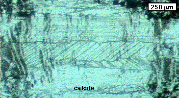
Fibrous:
-
High to extreme length/width ratio of grains (>10 ...
>100)
-
Fibrous shape not determined by crystal habit
-
Fibrous shape independent of crystallographic orientation
of grains
-
Shape of all grains identical
-
All fibres parallel
-
No nucleation during growth
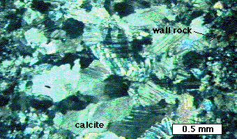
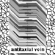
(Antitaxial calcite vein in carbonaceous shales, Arkaroola, South Australia) |
 |
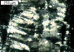
NB. A fibrous texture can be formed by fibrous
sub-grains or twins, while the true grains may not really be fibrous. Here
you see a section perpendicular to the fibres in an antitaxial vein. One
grain covers most of the image. The fibres are much smaller and are defined
by sub-grain and possibly twin-boundaries.
(Antitaxial calcite vein in carbonaceous shales, Arkaroola, South
Australia) |
 |
Elongate blocky:
-
Low to high length/width ratio of grains (<10)
-
Elongate shape not determined by crystal habit
-
Fibrous shape often related to crystallographic orientation
of grains
-
Not all grains have identical shape
-
Long axes of grains in approximately same direction
-
No nucleation during growth
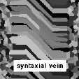
(Syntaxial/asymmetric quartz vein from Cape Liptrap, Victoria, Australia) |
 |
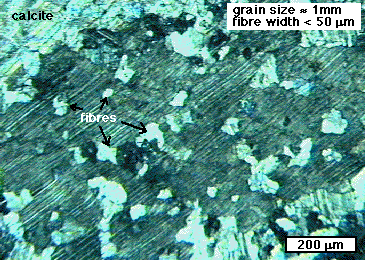
Blocky:
None of the specific characteristics of fibrous or elongate
blocky textures, in particular:
-
Often continuous nucleation
-
No elongate shape and/or shape preferred orientation
of crystals
(Calcite vein in carbonaceous shales, Arkaroola, South Australia) |
 |
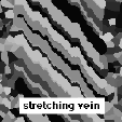
Stretched:
-
Elongate crystals
-
Parts of pre-existing grain at both ends of crystals
-
Often "radiator" structures and/or jogs on grain boundaries
(Calcite vein in carbonaceous silt stone and shales, Arkaroola, South
Australia) |
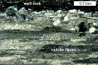 |
Slicken-fibres:
-
Fibrous or elongate blocky crystals
-
Long axis of crystals at low angle or parallel to vein
wall
(Calcite vein in carbonaceous shales, Arkaroola, South Australia) |
- Terms relating
to site(s) of precipitation during growth of a vein
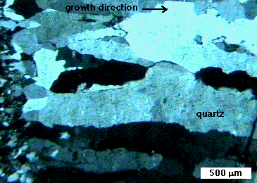
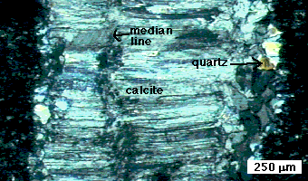
Syntaxial veins:
-
Growth persistently occurs at the same plane
-
Growth occurs in the middle of the vein
-
Growth often commences by syntaxial (crystallographic
sense) overgrowth of wall rock grains
-
Often elongate blocky
-
Oldest precipitate is on vein - wall rock contact; youngest
on median plane
-
Individual crystals do not extend across median plane
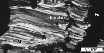
(Asymmetric antitaxial quartz vein from BIFs in Hammersley Ranges,
W.Australia) |
 |
Antitaxial veins:
-
Growth persistently occurs at the same planes
-
Growth occurs on the two vein - wall rock contact surfaces
-
Mineral(s) growing in the vein are often absent in,
or a minor constituent of the wall rock
-
Often fibrous
-
Youngest precipitate is on vein - wall rock contact;
oldest on median plane
-
Individual crystals extend across median plane
Ataxial or stretching
veins:
-
Growth occurs at various sites (cracks) over time
-
Mineral(s) growing in the vein are often major constituent
of the wall rock
-
Stretched crystals
-
-> No consistent variation from young to old precipitate
-
-> No median plane
-
-> Individual crystals extend from wall to wall of the
vein
Cracks can occur inside the vein only or they can occur
randomly (but usually parallel). In the second case, the vein contains
many slivers of wall-rock.
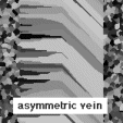
Asymmetric veins:
-
Growth commences by ataxial growth (usually, but can
be syn/antitaxial)
-
One side of the vein becomes preferred growth plane
-
Often elongate blocky
-
-> Veins are asymmetric
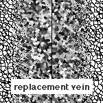
Replacement veins:
-
Vein precipitate is not in newly created space, but
replaces pre-existing minerals.
-
Usually vague edges
-
Inclusions of pre-existing grains often remain
-
Crystal shape often blocky or determined in shape and
size by pre-existing texture
 |
Composite veins:
-
Combination of syntaxial growth on both margins of vein
and syntaxial growth in centre of vein.
-
Usually different minerals forming syntaxial and antitaxial
parts
(Calcite + quartz vein in carbonaceous shales from Arkaroola, South
Australia) |
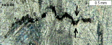
(Stylolite in calcite vein in carbonaceous shales from Arkaroola,
South Australia)
|
|
Stylolites:
Stylolites are in a way the opposite of veins (hence the term 'anti-crack'
which is sometimes used).
-
Continuous / repetative dissolution on a plane
-
Accumulation of insoluble material (often dark/opaque) on stylolite
-
Saw-teeth shape develops, due to differences in dissolution rate on either
side of stylolite
-
Saw-teeth indicate the direction of shortening
- Tracking of opening trajectory
The opening trajectory is the path that two, originally
adjacent, points on the opposite vein walls travelled relatively to each
other as the vein grew. Fibres & elongate blocky crystals often track
the opening trajectory, but not always completely (=partial tracking).
Ghost fibres may sometimes give a more reliable indication
of the opening trajectory than normal fibres. Ghost fibres are trails of
a different mineral growing off a specific point (grain) on the wall rock.
- Veins and structural analysis
The wide variety of internal structures of veins,
vein shapes and vein arrangements make veins useful structures for structural
analysis. Quite often the elongate blocky or fibrous crystals in a vein
allow us to determine the whole history of the formation of a vein, giving
insight in the deformation history of its host rock. The micro-structures
should of course be correctly interpreted (syntaxial or antitatxial, partial
or complete tracking, etc.).
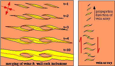
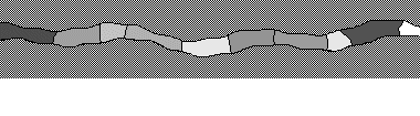
Veins also often form in arrays. The left image shows
a group of veins that originally formed at a small angle with the horizontal.
Interaction between the veins caused them to merge into one horizontal
vein with wall rock inclusions. The right image shows a set of sigmoidal
veins. The veins did not form all at the same time and are in different
stages of development (see film below). Sigmoidal vein arrays are often
useful kinematic indicators.
|
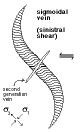
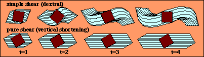
- Sigmoidal veins
Opening (widening) of a vein is in maximum instantaneous
stretching direction
-
opening trajectory records kinematics of deformation
Propagation of vein-tips parallel to maximum instantaneous
shortening direction
Opening of vein parallel to maximum instantaneous
extension direction
Vein rotates when deformation is non-coaxial
|
Strain/pressure
fringes & shadows around rigid objects
A rigid object (e.g. pyrite crystal) disturbs the
stress and strain field around it during deformation. On the sides of the
object normal to maximum compression, differential stress and pressure
are highest (high strain areas). On the sides of the object normal to minimum
compression, differential stress and pressure are lowest (low strain areas).
Difference in pressure can lead to material transport from strain cap to
pressure shadow or pressure fringe (alternatively called strain shadow
and strain fringe).
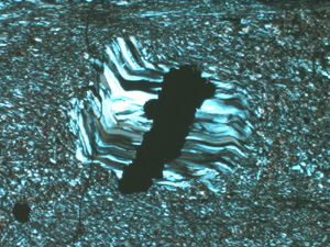
Pressure fringe of fibrous quartz around a concretion of iron ore in
a BIF-chert from the Hamersley ore province, Pilbara, West Australia.
Width of view 2.3 mm, crossed polars.
|
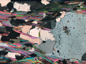
Quartz + mica pressure shadow adjacent to a quartz porphyroclast (on
right, grey grain with inclusions) in a quartz-mica schist from Nooldoonooldoona
Waterhole, S.W. Mount Painter Inlier, Arkaroola, South Australia. Width
of view 3.2 mm, crossed polars. Note the sharp boundary of the pressure
fringe, in contrast to the vague boundary of the pressure shadow
|
- Fringes versus shadows
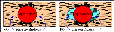
| distributed precipitation in low pressure area:
pressure shadow
-
usually blocky texture
-
non-distinct boundary of pressure shadow
-
similar to replacement veins
|
localised precipitation in low pressure area:
pressure fringe
-
usually fibrous or elongate blocky texture
-
sharp edge of pressure fringe
-
similar to syntaxial/antitaxial/composite veins
|
NOTE: later recrystallisation may produce a blocky
texture in a pressure fringe, making it look like a pressure shadow.
- Syntaxial
versus antitaxial fringes
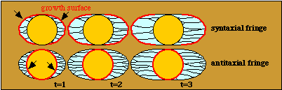
Syntaxial fringe:
-
precipitation is on outside of pressure fringe:
between
fringe & wall rock
-
precipitate can be same mineral as core object with
crystallographic continuity between object and fringe
-
relatively uncommon
|
Antitaxial fringe:
-
precipitation is on inside of pressure fringe:
between
object & fringe
-
precipitate usually different mineral as core object
-
relatively common (typically pyrite with quartz and/or
calcite fringe)
|
Notice that at the syn-/antitaxial terminology for veins and fringes
seems inconsistent. Reason:
-
in veins, the wall rock is reference material
-
in syntaxial veins crystals grow syntaxially from wall rock grains
-
in antitaxial veins crystals grow antitaxially towards wall rock
-
in fringes, the core object is reference material
-
in syntaxial fringes crystals grow syntaxially from core object
-
in antitaxial fringes crystals grow antitaxially towards core object
-
Displacement versus face-controlled growth
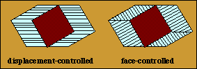
Displacement-controlled growth:
-
Growth direction of fringe crystals (fibres) = opening
direction
Face-controlled growth:
-
Growth direction of fringe crystals (fibres) = normal
to object surface
- Deforming versus non-deforming
fringes
The fringe mineral can be very strong compared to
the surrounding material and behave as rigid material. The texture in the
fringes is preserved. The fringe gets deformed if the fringe mineral is
not effectively rigid.
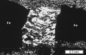 -
Pressure shadows/fringes in boudin necks
-
Pressure shadows/fringes in boudin necks
Pressure shadows and fringes also occur in boudin-necks of competent
layers or in between parts of broken rigid (but brittle) crystals.
(Quartz fringe in broken lump of iron ore in BIF from Hammersley
Ranges, Western Australia)
- Pressure shadows/fringes and structural analysis
As with veins, the shape of pressure shadows / fringes
and the internal texture of fringes often provide excellent information
about the kinetics of deformation during their formation:
-
degree of non-coaxiality of deformation
-
amount of deformation (finite strain)
-
possible changes in deformation kinetics
The crack-seal mechanism
The crack-seal mechanism (Ramsay
1980) is the favoured mechanism for about all veins with elongate crystals.
In this model growth occurs in many repeated small increments: crack-seal
events:
-
Crack event -> opening of narrow open crack, filled
with fluid
-
Seal event -> Precipitation fills (=seals) the crack
again
Most telling microstructural indicators of crack-seal
mechanism are regularly spaced trails of small inclusions (typically small
micas or pieces of wall rock or fluid inclusions). Opening per crack event
is generally in the order of 10 mm.
Elongate blocky and stretching textures are very
well explained by, and often show evidence of crack-seal growth (inclusions,
radiator structures)
- Crack-seal mechanism, pressure and fluid flow
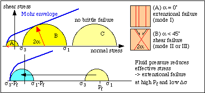
The crack-seal cycle involves the buildup of fluid pressure to enable fracturing (Crack). Then increased permeability allows fluid flow and material transport (Seal) and the fluid pressure drops.
- Presence of cracks -> fluid can flow through crack network
Presence of cracks -> brittle failure (often extensional)
Extensional failure -> high fluid pressure & low
differential stress
- Fibrous textures and the crack-seal mechanism:
some questions
How does material get to vein?
-
percolating fluid: precipitating in and clogging of cracks
-
diffusional transport: local material
Which textures develop ?
-
growth of crystals into open cracks is crystallographically
determined, resulting in elongate blocky textures and a preferred crystallographic
orientation
-
to get fibrous texture:
-
isotropic growth (special conditions, sub-grains or
twins)
-
growth is not in open crack, but on wall rock - fibre
tip contact (*)
Why often tracking of opening trajectory ?
-
tracking capability is a function of growth anisotropy
(crystallography), vein wall geometry (roughness) and crack width.
-
if crack width ->0 best tracking (*)
How to get symmetric opening in antitaxial vein ?
-
Simultaneous or alternating failure on both sides of
vein is difficult to imagine. More likely to see one side winning and get
asymmetric elongate blocky vein
-
Diffusional transport to and precipitation on surface
of vein gives symmetric antitaxial vein (*)
(*) These observations suggest that the crack
seal mechanism is not the only mechanism: it best explains the formation
of syntaxial or asymmetric elongate blocky veins. Fibrous
antitaxial veins may form without repeated cracking and sealing, but by
continuous growth on the surface of the vein, with diffusional transport
of material to the vein.